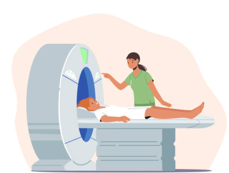
The diagnosis of Multiple Sclerosis is considered by physicians after patients report various symptoms including changes in vision, motor weakness, incoordination, change in walking, fatigue, and bowel and bladder issues. The evaluation almost always includes an MRI of the brain and spinal cord, and often also cerebral spinal fluid examination via a spinal tap and measurement of electrical impulses across the optic nerves.
One of the more common presentations of MS is cognitive impairment, including memory loss, which is often not recognized. In fact, 40-65% of all MS patients experience cognitive impairment, which can be one of the most debilitating symptoms of the disease. Even so, the cause of these cognitive changes has been largely elusive.
Multiple Sclerosis occurs when the outer layer of neurons, called myelin, are attacked by the immune system. This stripping of this myelin, called demyelination, is visualized on certain T2 MRI sequences. This damage is seen as bright white spots, called hyperintensities. Over time, the accumulation of greater numbers of white spots reflects structural and functional abnormalities. Clinically, the increasing burden of white matter lesions is manifested as more symptoms and greater disability.
A recently published study (Azzimonti, et al., 2023) investigated the association of MRI changes with cognitive loss. The investigators compared the brain MRI of patients with MS with patients with no known neurologic illness over a 40-month stretch of time. They focused on associations of the degree of cognitive dysfunction with the amount and location of white matter lesions, the microstructure of the white matter, measurements of the shrinking of areas of the brain of nerve fibers that are not covered by myelin, and resting state functional connectivity, called resting state fMRI. Resting-state fMRI evaluates interactions between different areas of the brain that occur in a resting or “task-negative state”.
Not surprisingly, the healthy group had no change in brain structure or functional connectivity. Interestingly, the 29% of MS patients with cognitive impairment had more gray matter atrophy in certain areas of the brain than the MS patients who were shown not to suffer from cognitive impairment. Further, MS patients with cognitive impairment demonstrated reduced functional connectivity in the memory center and MS patients without cognitive impairment had increased functional connectivity in the left insula as compared to those cognitively unaffected. Most importantly, there had not been increased white matter lesions or microstructural changes in either MS group over this period of time. The hope is that by identifying the areas involved in cognitive decline in MS patients, therapies can be targeted to these areas to preserve cognition in these patients.
Disease-modifying therapies can help reduce the number of MS attacks on the nervous system and ultimately reduce many forms of disability associated with disease progression. Many manifestations of disease progression can be insidious and significant worsening may not be detected by patients or physicians on a timely basis. This is especially true for cognitive impairment.
BeCare MS link can help detect changes in the functioning of the central nervous system earlier because it delivers quantified neurologic measurements augmented by AI analysis. BeCare MS Link offers established neuropsychiatric testing to assess cognitive impairment.
Reference:
Azzimonti, M., Preziosa, P., Pagani, E. et al. Functional and structural brain MRI changes associated with cognitive worsening in multiple sclerosis: a 3-year longitudinal study. J Neurol 270, 4296–4308 (2023). https://doi.org/10.1007/s00415-023-11778-z
Please send details about testing link